Objective
The Canine/Premolar contact is difficult to capture. Parallel technique is hard to achieve even with film, because of the curvature of anatomy targeted in a bitewing. With sensors, which are not flexible, the challenge is even greater. Indeed, most sensors on the market make matters worse by having a mesial dead space larger than a film packet’s. Therefore, design a sensor with mesial dead space approximately the same as a film packet’s mesial dead space.
Design
For historical reasons, most sensors place necessary electronics near the edge where the cord exits the sensor. The result is a dead space of up to 6mm, since the electronics cannot also be part of a sensor’s active imaging area. So, for bitewing purposes, the very edge used to capture the canine/premolar contact may not capture any image at all for its first 3-6mm. Clinicians who regularly place sensors instantly see this as a problem.
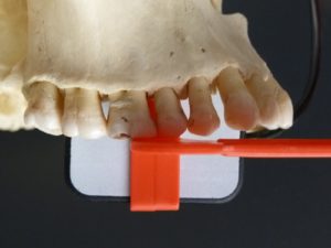
XDR clinicians asked for the dead space to be minimized. By moving the electronics elsewhere, XDR engineers achieved a 2mm mesial dead space, little more than the dead space common with film packets. Figure 1 shows how active imaging area at the mesial edge can help capture of the canine/premolar contact.
|
|
Results
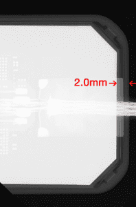
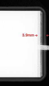
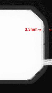
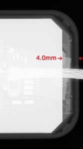
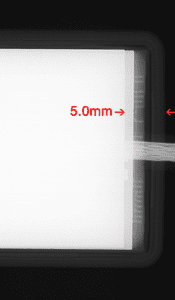
Figures 2a through 2d demonstrate how XDR’s imaging area extends further toward the sensor’s mesial edge. XDR has patented this approach under US #9357972B .
 |
 |
 |
 |
 |
Conclusion
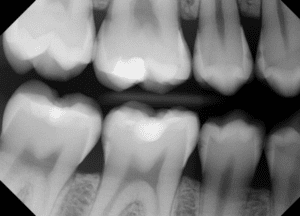
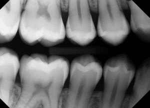
Doctors report that good parallel technique is easier to achieve with XDR’s Anatomic sensor. The maximized mesial imaging area aids capture of the canine/premolar contact. And that bitewing images more often display 8 contacts for diagnosis (see figures 3a and 3b).
|
|
|
|
 Bernice Ko, DDS