Objective
The quality of a radiographic system is only as good as its ability to capture the desired anatomy. For instance, if the clinician has cannot capture the canine/premolar contact because of poor sensor design, then even excellent resolution is pointless.
So:
- Is it easy to see if the sensor is placed properly?
- When optimally placed, does the active image area cover the targeted anatomy?
Design
Intraoral Visibility. Careful placement and orientation of the sensor are important, whether pursuing interproximal contacts or third-molar apices. XDR found that making the active surface white – without patterns or logos – made the sensor easier to see in the darkness of a mouth.
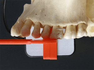
Canine/Premolar Contact. Even when optimally placed, excessive dead space along the mesial edge of the sensor makes it difficult to capture certain anatomy, such as the canine-premolar contact. In XDR’s patented design (US #9357972B2), XDR has decreased this dead space, maximizing the imaging area along the mesial edge.
 |
 |
|
|

Results
Ergonomic Features
| Objective | Feature |
| Canine-Premolar Contact | Maximal Mesial Imaging Area |
| Intraoral Visibility | White Face |
| Comfort | Rounded Corners |
| Comfort | Beveled Corners |
| Ease of Placement | Thin |
| Ease of Placement | Small Button |
| Ease of Cleaning | No Creases or Crevices |
| Sterilizability | Immersible (Waterproof) |
| Strong Cord | Kevlar Cable |
| Shock Resistance | Protection Plate |
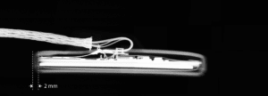
| Handling Ease | Two-Meter Cord |
Conclusion
Many sensors provide some of these features. Only the XDR Anatomic Sensor provides all of them.